
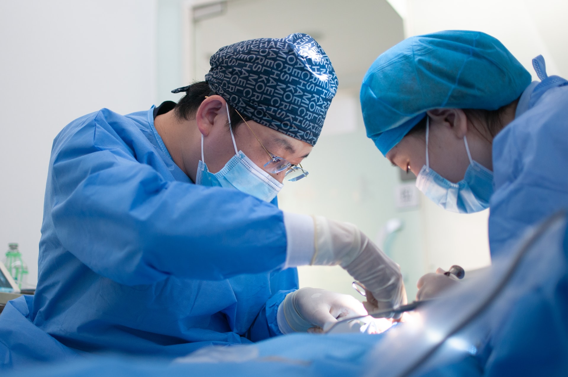
The Israeli Galilee Medical Center (GMC) saw yet another innovation in the medical field through amalgamation with contemporary technology as its surgeons combined 3D printing and augmented reality (AR) to treat an eye socket fracture patient.
Using 3D printing, they fabricated a metal implant and then used Microsoft HoloLens to precisely put it within the patient’s skull who was ailing due to a face injury. The inclusion of AR helped them to execute the procedure swiftly with a higher degree of accuracy, leading to an enhanced surgical outcome and eliminating the need for follow-up surgery.
“Utilizing a 3D printer and AR resulted in both a particularly accurate execution of the operation and a significant reduction in time. This technology will contribute to improved clinical outcomes and reduce repeated imaging and surgeries,” said Professor Samer Srouji who led the procedure.
The usage of 3D printing to create a face implant is already a popular concept across the world with multiple health organizations putting it to use regularly for procedures. The UK’s NHS has been employing the technology for facial reconstructions for over four years. Texas A&M University has also experimented with stem cell-based implants that enhance cranial regeneration.
Even the HoloLens was first introduced in 2016 and multiple developers have released software that integrates the device with 3D modeling equipment. Therefore, the standalone technologies are not really new but their combination in such an environment for this purpose presents a lot of opportunities for their usage in the medical field.
The amalgamation was a result of the surgeons’ effort to treat the 31-year-old man with a fractured left eye socket without impairing the aesthetics of his eyes. They collaborated with their counterparts at the Sheba Medical Center (SMC) to develop the novel method for executing the surgery successfully.
The surgeons took input from the CT scans of the patient to reconstruct the shape of the floor of his eye socket accurately. They 3D printed a customized titanium graft by first projecting the healthy side onto the damaged part to build a complete 3D model of the patient’s skull.
A computer program containing both a model of the patients’ skull and the titanium graft guided the surgeon wearing the AR glasses to accurately place the 3D printed metal plate over the damaged part. The innovative procedure enabled them to bring down the time required for surgery down to just an hour and a half.
Srouji accredits the success of the initiative to the continued development of the GMC’s 3D Point of Care unit despite the ongoing pandemic. Both GMC and SMC had carried on with the experimentation of 3D technologies and further aim to discover more applications for them for several surgical procedures in the time to come.
Biocompatible implants, that help patients to heal better, have also started garnering attention from scientists across the globe with the increasing popularity of 3D printing.
Scientists at the Skolovo Institute of Science and Technology (Skoltech) have developed a simulation-based approach through 3D printing of personalized ceramic bone grafts. They have managed to create designs with larger pores that make the process of the graft’s fusion with the organic tissues simpler.
Their counterparts at the Dutch Delft University of Technology have devised a novel method to treat critical bone defects through 3D printed biodegradable magnesium scaffolds. These grafts stimulate bone regeneration and then degrade naturally inside the body over time.
Not depriving the animal kingdom of the advancements in medical technology, the scientists at the Animal Health Trust (AHT) and the University of East Anglia (UEA) have come up with a way to support bone regeneration in horses through 3D printed scaffolds. They have potentially transformed the way veterinary fracture repair surgeries are conducted by devising a way to create novel osteoblasts by injecting stem cells into their additive devices.
Follow us on LinkedIn
Read other Articles